SAY NO TO DRUGS

Saturday, October 30, 2010

Trabeculectomía

En oftalmología, la trabeculectomía es un procedimiento quirúrgico aplicado en el tratamiento del glaucoma para aliviar la presión intraocular quitando parte de la red trabecular del ojo y estructuras adyacentes. Es la cirugía más común para el tratamiento del glaucoma. La trabeculectomía permite el drenaje del humor acuoso desde dentro del ojo hacia la parte inferior de la conjuntiva donde es absorbida.

Este procedimiento se lo realiza comunmente bajo anestesia monitoreada utilizando un block retrobular o peribulbar, o una combinación de anestesia topica y subtenoniana. Para llevar a cabo una trabeculectomía se realiza una perforación en la porción más externa del ojo o esclerótica, hasta llegar a un espacio del interior del órgano de la visión que se llama cámara anterior. Por este nuevo conducto creado por la cirugía se consigue que drene hacia el exterior el humor acuoso.


Trabeculectomía realizada por el Dr Rodrigo Rodriguez

Online Schools of Nursing - Getting A Nursing Degree

Earning a nursing degree from online schools of nursing is becoming more and more popular. The flexibility and versatility these online nursing schools offer poses a lot of benefits.


Those looking to get a nursing degree can definitely take advantage of all the benefits online schools of nursing have to offer.

When earning a nursing degree online, you basically complete the non-clinical courses because clinical and laboratory classes
must be completed in person at a medical facility also operated by the online school of nursing. It is important to know that there are no online nursing programs that allow you to fully complete the degree online. Nevertheless, it is one good way to become a registered or practical nurse.
To find online nursing schools such as online practical nursing schools, you need to do a little research online. There are many websites offering online nursing courses. In fact, even online nursing PHD programs are available on the Internet. On Google or even on other reliable search engines, type the keywords that will help you find the online nursing schools you would like to attend. At the very least, you can type the words "online nursing degree" or "nursing degree online". You may also type the name of your city or state that you would like your nursing program to be in. The search engine will show you results with links to many online nursing schools. Check out some of these sites to see in detail what they offer and find out if their offerings match what you are looking for. There are some things that you consider when choosing the online school to attend to.

First and foremost, find out if the nursing program allows you to work at your own pace or the one that follows the school's schedule. A program that allows you to work at your own pace will allow you to finish or complete the work at a schedule or pace that suits you. However, there are many nursing programs structured like a traditional school or college. You should also find out the date when you can start. Some online schools for nursing have rolling enrollment which means that you can start at any date you prefer. Others designate a starting date.

You should also check if the online school has an academic advisor. It is a good idea to settle for one that assigns an academic advisor for each student because the advisor will help and work with students closely to help in planning out the course of study. An academic advisor also helps students keep on tract, connects with the students online, and coordinates with the students' clinical classes.

You should also check the costs of the nursing program because not all schools have the same charges. A thorough search of programs will help you land on the most affordable yet right online school for you. It is also good if you can find out if the school offers financial assistance. Many online nursing programs offer assistance and it is a good idea to take advantage of this financial help.

Once you have completed your researchBusiness Management Articles, you can narrow your choices of online schools of nursing down to a few until you land on the best one.

Friday, October 29, 2010

Red Trabecular

La red trabecular es un área de tejidos esponjosos ubicados alrededor de la base de la córnea, cerca del cuerpo ciliar. La función de la misma es el drenaje del humor acuoso desde la cámara anterior del ojo hacia el Canal de Schlemm que finalmente desembocan en el sistema sanguíneo. El tejido esponjoso que forma la red trabecular está recubierto por células especiales llamadas trabeculocitos.

La red trabecular está compuesta de tres partes: 1) la red uveal interna, que es la más cercana al ángulo de la cámara anterior y que contienen trabéculas o tabiques orientados en forma radial que encierran pequeños espacios internos; 2) red corneoescleral, la cual tiene gran cantidad de elastina (proteina con funciones estructurales) en forma de láminas delgadas perforadas; 3) tejido yuxtacanalicular, que yace en forma adyacente al canal de Schlemm y está compuesto de tejido conectivo.

La red trabecular desempeña un papel importante, ya que evita el aumento anormal de la presión intraocular, lo que puede provocar un glaucoma. Esto se debe que a través de ella fluye el humor acuoso hasta que finalmente es drenado al sistema venoso por el Canal de Schlemm. Cuando la red trabecular no cumple esta función de drenaje, se produce un aumento de la presión intraocular del humor acuoso.

How To Get The Advanced Practical Nursing Job You Want (Ebook Killer Version)

How To Get The Advanced Practical Nursing Job You Want (Ebook Killer Version)

How To Get The Advanced Practical Nursing Job You Want (Ebook Killer Version)


Product Description

Do you know what it takes to get the job you want? In this ebook, the original
“How To Get A Job You Want And Succeed Once You’re There,” has been
edited only for those interested this particular job. This ebook contains helpful
tips for keeping a job, interviewing, a resume template all ready to go, and
very comprehensive information regarding the career field from job growth, to
educational requirements, to the very nature of the job, and everything in
between. Get the job you want with the information in this ebook!

THIS EBOOK HAS BEEN FORMATTED INTO A SIMPLIFIED VERSION SO IT
IS NOW COMPATIBLE WITH SMARTPHONES. GRAPHICS HAVE BEEN
OMITTED FOR A SIMPLIFIED TEXT READING EXPERIENCE.

(EBOOK KILLER VERSION INFORMATION) Ever notice that most ebooks
have basically the same information? Hell, even sometimes they have the
same information word-for-word? That's because all of those ebooks use the
same couple of sources for their information, and all they do is re-write a
couple or articles or chapers, and then click "publish." With an "Ebook Killer
Version," we're giving you all of the original, unedited information used to
create those ebooks so you don't have to keep buying the same stuff over and
over again. We're going to give you everything, even if some of the chapters
seem like duplicates, so you can save your money.


Click Here How To Get The Advanced Practical Nursing Job You Want (Ebook Killer Version)

Maternal Child Nursing Care - Text and E-Book Package

Maternal Child Nursing Care - Text and E-Book Package

Maternal Child Nursing Care - Text and E-Book Package

Maternal Child Nursing Care - Text and E-Book Package

Product Description

Evolve eBook

The Evolve eBook gives you electronic access to all textbook content with plenty of added functionality. Not only can you search your entire library of eBooks with a single keyword, you can create your own customized study tool by highlighting key passages, taking and sharing notes, and organizing study materials into folders. Add additional eBooks to your collection to create an integrated digital library! Your Evolve eBooks are conveniently accessible either from your hard drive or online.


Book Description

This market-leading textbook provides just the "right amount" of maternity and pediatric content in an easy-to-understand manner. Divided into two sections, the first part of the book includes 28 chapters on maternity nursing and the second part contains 27 chapters covering pediatric nursing. Numerous illustrations, photos, boxes, and tables clarify key content and help you quickly find essential information. And because it's written by market-leading experts in maternity and pediatric nursing, you can be sure you're getting the accurate, practical information you need to succeed in the classroom, the clinical setting, and on the NCLEX® examination.

Nursing Diagnosis Handbook: An Evidence-Based Guide to Planning Care

Nursing Diagnosis Handbook: An Evidence-Based Guide to Planning Care

Nursing Diagnosis Handbook: An Evidence-Based Guide to Planning Care

Book Description

Write individualized care plans with ease and confidence!

Product Description

Use this convenient resource to formulate nursing diagnoses and create individualized care plans! Updated with the most recent NANDA-I approved nursing diagnoses, Nursing Diagnosis Handbook: An Evidence-Based Guide to Planning Care, 9th Edition shows you how to build customized care plans using a three-step process: assess, diagnose, and plan care. It includes suggested nursing diagnoses for over 1,300 client symptoms, medical and psychiatric diagnoses, diagnostik procedures, surgical interventions, and clinical states. Authors Elizabeth Ackley and Gail Ladwig use Nursing Outcomes Classification (NOC) and Nursing Interventions Classification (NIC) information to guide you in creating care plans that include desired outcomes, interventions, patient teaching, and evidence-based rationales.

  • Promotes evidence-based interventions and rationales by including recent or classic research that supports the use of each intervention.
  • Unique! Provides care plans for every NANDA-I approved nursing diagnosis.
  • Includes step-by-step instructions on how to use the Guide to Nursing Diagnoses and Guide to Planning Care sections to create a unique, individualized plan of care.
  • Includes pediatric, geriatric, multicultural, and home care interventions as necessary for plans of care.
  • Includes examples of and suggested NIC interventions and NOC outcomes in each care plan.
  • Allows quick access to specific symptoms and nursing diagnoses with alphabetical thumb tabs.
  • Unique! Includes a Care Plan Constructor on the companion Evolve website for hands-on practice in creating customized plans of care.
  • Includes the new 2009-2011 NANDA-I approved nursing diagnoses including 21 new and 8 revised diagnoses.
  • Illustrates the Problem-Etiology-Symptom format with an easy-to-follow, colored-coded box to help you in formulating diagnostic statements.
  • Explains the difference between the three types of nursing diagnoses.
  • Expands information explaining the difference between actual and potential problems in performing an assessment.
  • Adds detailed information on the multidisciplinary and collaborative aspect of nursing and how it affects care planning.
  • Shows how care planning is used in everyday nursing practice to provide effective nursing care.

Nursing Diagnosis Handbook: An Evidence-Based Guide to Planning Care

Mesothelioma Treatment Part 2

Treatment and medicine
What treatment you undergo for mesothelioma depends on your health and certain aspects of your cancer, such as its stage and location. Unfortunately, mesothelioma often is an aggressive disease and for most people a cure isn't possible. Mesothelioma is usually diagnosed at an advanced stage — when it isn't possible to remove the cancer through an operation. Instead, your doctor may work to control your cancer to make you more comfortable.
Discuss treatment goals with your doctor. Some people want to do everything they can to treat their cancer, even if that means enduring side effects for a small chance of an improvement. Others prefer treatments that make them comfortable so that they can live their remaining time as symptom-free as possible.
Surgery
Surgeons work to remove mesothelioma in instances where it is diagnosed at an early stage. Sometimes it isn't possible to remove all of the cancer. In those cases, surgery may help to reduce the signs and symptoms caused by mesothelioma spreading in your body. Surgical options may include:
      Surgery to decrease fluid buildup. Pleural mesothelioma may cause fluid to build up in your chest, causing difficulty breathing. Surgeons insert a tube or catheter into your chest to drain the fluid. Doctors may also inject medicine into your chest to prevent fluid from returning (pleurodesis).
      Surgery to remove the tissue around the lungs or abdomen.Surgeons may remove the tissue lining the ribs and the lungs (pleurectomy) or the tissue lining the abdominal cavity (peritonectomy) in order to relieve signs and symptoms of mesothelioma.
      Surgery to remove as much of the cancer as possible (debulking). If all of the cancer can't be removed, surgeons may attempt to remove as much as possible.
      Surgery to remove a lung and the surrounding tissue.Removing the affected lung and the tissue that surrounds it may relieve signs and symptoms of pleural mesothelioma. If you'll be receiving radiation therapy to the chest after surgery, this procedure also allows doctors to use higher doses, since they won't need to worry about protecting your lung from damaging radiation.
Chemotherapy
Chemotherapy uses chemicals to kill cancer cells. Systemic chemotherapy travels throughout the body and may shrink or slow the growth of a pleural mesothelioma that can't be removed using surgery. Chemotherapy may also be used before surgery (neoadjuvant chemotherapy) to make an operation easier or after surgery (adjuvant chemotherapy) to reduce the chance that cancer will return.
Chemotherapy drugs may also be heated and administered directly into the abdominal cavity (intraperitoneal chemotherapy), in the case of peritoneal mesothelioma, or into the chest cavity (intrapleural chemotherapy), in the case of pleural mesothelioma. Using this strategy, chemotherapy drugs can reach the mesothelioma directly without injuring healthy cells in other parts of the body. This allows doctors to administer higher doses of chemotherapy drugs.
Radiation therapy
Radiation therapy focuses high-energy beams, such as X-rays, to a specific spot or spots on your body. Radiation may reduce signs and symptoms in people with pleural mesothelioma. Radiation therapy is sometimes used after biopsy or surgery to prevent mesothelioma from spreading to the surgical incision.
Combination therapy
Surgery, chemotherapy and radiation therapy may be used in various combinations to treat both pleural mesothelioma and peritoneal mesothelioma.
Clinical trials
Clinical trials are studies of new mesothelioma treatment methods. People with mesothelioma may opt for a clinical trial for a chance to try new types of treatment. However, a cure isn't guaranteed. Carefully consider your treatment options and talk to your doctor about what clinical trials are open to you. Your participation in a clinical trial may help doctors better understand how to treat mesothelioma in the future.
Clinical trials are currently investigating a number of targeted drugs. Targeted drug therapy uses drugs to attack specific abnormalities within cancer cells. Targets being studied in mesothelioma include a substance that cancer cells make to attract new blood vessels that bring oxygen and nutrients to the cancer. Researchers hope drugs that target these areas can help kill mesothelioma cells.
Treatment for other types of mesothelioma
Pericardial mesothelioma and mesothelioma of tunica vaginalis are very rare. Early-stage cancer may be removed through surgery. Doctors have yet to determine the best way to treat later stage cancers, though. Your doctor may recommend other treatments to improve your quality of life.
Alternative Treatment
Mesothelioma can cause pressure within your chest that can make you feel as if you're always short of breath. Breathlessness can be distressing. Your doctor may recommend using supplemental oxygen or taking medications to make you more comfortable, but often these aren't enough. Combining your doctor's recommended treatments with complementary and alternative approaches may help you feel better.
Alternative treatments that have shown some promise in helping people cope with breathlessness include:
  •       Acupuncture. Acupuncture uses thin needles inserted at precise points into your skin.
  •       Breath training. A nurse or physical therapist can teach you breathing techniques to use when you feel breathless. Sometimes you may feel breathless and begin to panic. Implementing these techniques may help you feel more in control of your breathing.
  •      Relaxation exercises. Slowly tensing and relaxing different muscle groups may help you feel more at ease and breathe easier. Your doctor may refer you to a therapist who can teach you relaxation exercises so that you can do them on your own.
  •      Sitting near a fan. Directing a fan to your face may help ease the sensation of breathlessness.

Thursday, October 28, 2010

The Mental Status Exam

The mental status exam, is an assessment tool that helps identify psychological symptoms that may assist the practitioner determine if there is a psychogenic problem. When assessing mental status, it is important to adjust questions and categories to avoid age and/or cultural bias.
Category Description
Appearance General appearance, grooming and gait. This is best observed as the client comes into the room. Grooming is one of the earliest areas to deteriorate when mental function has diminished.
Behavior Speech, eye contact, body language, response to the environment. Observe for appropriate use of personal space. Does the person come right into your face, or stand an unusual distance away.
Insight The ability of the client to be aware of one’s own abilities. The ability to analyze a problem objectively. Ask the client to explain a problem.
Intellectual Functioning Simple calculations, ability to abstract or think symbolically and categories of association. This is done through direct questioning using math, proverbs or analogy.
Judgment Assesses decision-making abilities. Ask client What he would do in a dilemma regarding an important decision.
Memory Immediate recall, recent memory, remote memory. Ask the client about a recent current event that both you and the client should know. Ask about some event in the past that should be known by both. Be very careful in this area to avoid cultural bias.
Mood and Affect Mood relates to the emotions of the moment while affects refers to the range of emotions displayed such as happy, sad, or unchanging. Compare in relation the client’s probable everyday behavior.
Orientation Assess for awareness of person, time, place, and purpose.
Perceptual Processes Awareness of self and one’s thoughts, reality and fantasy. Ask about delusions, illusions and hallucinations. Do not hesitate do ask direct questions.
Sensorium Ability to concentrate, perception of stimuli.
Thought Contents This assesses themes in conversation and is assessed by observing what the client discusses spontaneously in conversation.
Thought Processes This measures a stream of conscious or mental activity as indicated in speech. Observe for rate, flow, and ability to pursue a topic logically.


www.accessce.com

Neurological Assessment : Checks Pupils

Neurological Assessment : Checks Pupils


Neurological Assessment : Checks Pupils


  1. Observes Both Pupils Simultaneously For: Equality, Size and Shape.
    • Compares pupils for equality.
    • Determines size, dilated, constricted, pinpoint.
    • Determines irregularities in shape.

  2. Observes Direct Pupillary Light Reflexes.
    • Checks one pupil at a time.
    • Shines flashlight into eye from side.
    • Repeat other eye.

  3. Observes Consensual Pupillary Reflex
    • Shines flashlight into each eye alternately.
    • Observes opposite pupil. Opposite pupil should constrict when light shore.
    • Charts description of pupils: Equality, size, shape, reaction to light.

  4. Observes pupillary response to accommodation
    • Have patient follow a closer moving object such as a pen.
    • Pupils will constrict (or accommodate) to the closer moving object. * cannot be tested on blind or confused persons.

  5. Observes Extraocular Movements
    • Asks patient to focus on object.
    • Moves object; medical, lateral, superior, inferior and circular. In the pattern of an "H."
    • Observes movement of both eyes in each of above directions; notes abnormalities or weakness. A. Charts extraocular movements as "full" if no abnormalities or "unable to move eyes laterally, medially etc."
accessce.com

Presión intraocular

La presión intraocular es la presión que ejerce los fluidos del humor acuoso dentro del globo ocular. Está determinada por el equilibrio entre la cantidad de humor acuoso segregada por los procesos ciliares y la cantidad de este fluido que se drena a través de la red trabecular que se encuentra en el ángula de la cámara anterior del ojo. La presión intraocular se la mide con un instrumento llamado tonometro.

En la parte anterior del ojo, entre el cristalino y la córnea, se encuentra el humor acuoso. El humor acuoso ocupa el 3% del interior del ojo, se renueva constantemente, la producción y reabsorción son continuas y se mantienen equilibradas en circunstancias normales. Si se produce un desequilibrio por exceso de producción o bloqueo en la reabsorción, la presión intraocular se eleva por encima de las cifras adecuadas para el correcto funcionamiento del ojo.

El valor medio de la presión intraocular es 16 mm de Hg y puede medirse fácilmente con ayuda de un dispositivo que se llama tonómetro. El equilibrio entre producción y reabsorción del humor acuoso es el principal factor que determina el nivel de presión intraocular. Por otra parte la elevación de la presión intraocular o hipertensión ocular es el principal factor de riesgo para que se desarrolle una enfermedad del ojo conocida como glaucoma.

Wednesday, October 27, 2010

Vital Signs - Blood Pressure

Blood Pressure

Vital Signs - Blood Pressure


Blood Pressure : Pressure of blood against the walls of the arteries

Systolic : Number that is on the top, and when heart is contracting

Diastolic : Number that is on the bottom, and when heart is at rest

Systolic range : 90 - 140

Diastolic range : 60 - 90

Hypertension : High blood pressure, above 140 systolic or over 90 diastolic

Hypotension : Low blood pressure, under 90 over 60

To measure systolic : Sound of first beat

To measure diastolic : No beat is heard

Hypertension thickens heart muscle (hypertrophy) and reduces chamber in size

Thigh cuff for large arms, Small cuff pediatrics

Sphygmomanometer is instrument use to take blood pressure

Pulse pressure: Difference between systolic and diastolic

Vital Signs - Pulse

Pulse

Vital Signs - Pulse


Rate is : Number of beats per minute

Rhythm is : Regularity of beats

Normal range of adults : 60 - 100 per minute

Pulse : Can be weak, bounding or absent for short period of time

Rhythm : Can be regular or irregular

Palpate for : Rhythm, rate, and strength

Optimal finding : 80 per min. strong, and reg.

Tachycardia : Over 100 beats per minute

Bradycardia : Under 60 beats per minute

To measure pulse : count 30 sec.X 2

For irregular pulse : count the full 60seconds

Auscultate : Use stethoscope

Pulse deficit : Difference of apical and radial.

Vital Signs Respirations

Respirations


Vital Signs Respirations


Respirations : How many breaths per minute

Adults: 12 - 20 / Infant slightly higher 20 - 40

Inhalation and Exhalation equals: 1 breath

To count breaths: Count 30 seconds by 2

Look for : Rhythm, rate, depth, and quality

Bradypnea: Under 12 breaths

Tachypnea: Over 20 breaths

Eupnea: Normal rate and depth

Apnea: Not breathing maybe 30 seconds or at all

Dyspnea: Difficulty in breathing

Orthopnea: Over bedside 90o postural position

Hyperpnea: Fast respirations

Cheyne Stokes: Increasing in rate and depth then periods of apnea - cyclic.

Kussmaul: Metabolic acidosis,usually the Diabetic. Rapid, very deep respirations intended to blow off carbondioxide.

Vital Signs - Temperature

Temperature


Vital Signs - Temperature


Oral - mouth

Time period 3 minutes
Normal range: 97.6 - 99.6 degrees
Absolute: 98.6 degrees

Rectal - Anus
Time period 3 minutes
Position -Lateral Sims
Normal range: 98.6 - 100.6 degrees
Absolute: 99.6 degrees

Axillary - Armpit
Time period 10 minutes
Normal range: 96.6 - 98.6
Absolute: 97.6 degrees
Otic or Tympanic Time period 10 sec. or less

Degree range is calibrated to rectal or oral
Hypothermia - Low body temperature
Hyperthermia - High body temperature
Pyrexia - High fever
Febrile - High fever
Afebrile - No fever

Things that can effect temperature: smoking, fluids, oxygen use, food, colds, or flu.(www.accessce.com)

Apgar Score

Apgar Scoring


Sign
0 points
1 point
2 points
A
Activity
(Muscle tone)
limp
limbs flexed
active movement
P
Pulse
(heart rate)
absent
<>
> 100 /min
G
Grimace
(response to smell or foot slap)
absent
grimace
cough or sneeze (nose)
cry and withdrawal of foot (foot slap)
A
Appearance
(color)
blue
body pink
extremities blue
black over
R
Respiration
(breathing)
absent
irregular
weak crying
good strong cry

The total Apgar score is the sum of the scores for the five signs.


Vitrectomía

En oftalmología, la vitrectomía es un procedimiento quirúrgico para remover parte o todo el humor vítreo del ojo, sustituyéndolo con líquido o gas. La vitrectomía es utilizada para el tratamiento de diferentes enfermedades oculares, como el desprendimiento de retina, la hemorragia vítrea y el agujero macular.

La vitrectomía anterior implica quitar pequeñas cantidades del humor vítreo de las estructuras frontales, mientras que la vitrectomía pars plana son cirugías de la parte más profunda del ojo también destinada a remover parte o todo el humor vítreo. Este último procedimiento por lo general se aplica en casos severos de miodesopsias.

Video de vitrectomía realizado por el Dr. Luciano Berretta

The 12 Cranial Nerves

The 12 Cranial Nerves


There are 12 pairs of cranial nerves. These nerves arise from the brain and brain stem, carrying motor and or sensory information.


Cranial nerve I : Olfactory nerve

The olfactory nerve is composed of axons from the olfactory receptors in the nasal sensory epithelium. It carries olfactory information (sense of smell) to the olfactory bulb of the brain. This is a pure sensory nerve fiber.

Cranial nerve II: Optic nerve

The optic nerve is composed of axons of the ganglion cells in the eye. It carries visual information to the brain. This is a pure sensory nerve fiber. This nerve travels posteromedially from the eye, exiting the orbit at the optic canal in the lesser wing of the sphenoid bone. The optic nerves join each other in the middle cranial fossa to form the optic chiasm.


Cranial nerve III: Oculomotor nerve

The oculomotor nerve is composed of motor axons coming from the oculomotor nucleus and the edinger-westphal nucleus in the rostral midbrain located at the superior colliculus level. This is a pure motor nerve. It provides somatic motor innervation to four of the extrinsic eye muscles: the superior rectus, inferior rectus, medial rectus, and the inferior oblique muscles. It also innervates the muscles of the upper eyelid and the intrinsic eye muscles (the pupillary eye muscle.) Together, CN III, CN IV and CN VI control the six muscles of the eye.


Cranial nerve IV: Trochlear nerve

The trochlear nerve provides somatic motor innervation to the superior oblique eye muscle. This cranial nerve originates at the trochlear nucleus located in the tegmentum of the midbrain at the inferior colliculus level and exits the posterior side of the brainstem. It is also a pure motor nerve fiber.


Cranial nerve V: Trigeminal nerve

The trigeminal is the largest cranial nerve . It provides sensory information from the face, forehead, nasal cavity, tongue, gums and teeth (touch, and temperature) and provides somatic motor innervation to the muscles of mastication or “chewing”.

This cranial nerve has 3 branches: the ophthalmic, maxillary and mandibular branches.

It is composed of both sensory and motor axons. The sensory fibers are located in the trigeminal ganglion and the motor fibers project from nuclei in the pons.


Cranial nerve VI: Abducens nerve

The abducens nerve carries somatic motor innervation to one of the extrinsic eye muscles, the lateral rectus muscle. It is another pure motor nerve fiber and originates from the abducens nucleus located in the caudal pons at the facial colliculus level.


Cranial nerve VII: Facial nerve

The facial nerve carries somatic motor innervation to the many muscles for facial expression. It carries sensory information form the face (deep pressure sensation) and taste information from the anterior two thirds of the tongue. It arises at the pons in the brainstem and it emerges through openings in the temporal bone and stylomastoid foramen and has many branches. It is composed of both sensory and motor axons.


Cranial nerve VIII: Vestibulocochlear nerve

The vestibulocochlear nerve innervates the hair cell receptors of the inner ear. It carries vestibular information to the brain from the semicircular canals, utricle, and saccule providing the sense of balance. It also carries information from the cochlea providing the sense of hearing. This cranial nerve branches into the Vestibular branch (balance) and the cochlear branch (hearing). The cochlear fibers originate from the spiral ganglion. It is pure sensory nerve fiber.


Cranial nerve IX: Glossopharyngeal nerve

The glossopharyngeal nerve innervates the pharynx (upper part of the throat), the soft palate and the posterior one-third of the tongue. It carries sensory information (touch, temperature, and pressure) from the pharynx and soft palate. It carries taste sensation from the taste buds on the posterior one third of the tongue. It provides somatic motor innervation to the throat muscles involved in swallowing. It provides visceral motor innervation to the salivary glands. This cranial nerve also supplies the carotid sinus and reflex control to the heart . It is composed of both sensory and motor axons and originates from the nucleus ambiguous in the reticular formation of the medulla.


Cranial nerve X: Vagus nerve

The vagus nerve consists of many rootlets that come off of the brainstem just behind the glossopharyngeal nerve. The branchial motor component originates from the nucleus ambiguous in the reticular formation of the medulla. The visceral component originates from the dorsal motor nucleus of the vagus located in the floor of the fourth ventricle in the rostral medulla and in the central grey matt er of the caudal medulla. It is the longest cranial nerve
innervating many structures in the throat, including the muscles of the vocal cords, thorax and abdominal cavity. It provides sensory information (touch, temperature and pressure) from the external auditory meatus (ear canal) and a portion of the external ear. It carries taste sensation from taste buds in the pharynx. It also provides sensory information from the esophagus, respiratory tract, and abdominal viscera (stomach, intestines, liver, etc.). It provides visceral motor innervation to the heart, stomach, intestines, and gallbladder. It is part of the ANS, the parasympathetic branch. It is composed of both sensory and motor axons. Other parasympathetic ganglia include CN III , CN VII and CN IX .


Cranial nerve XI: Spinal Accessory nerve

The spinal accessory nerve has two branches. The cranial branch provides somatic motor innervation to some of the muscles in the throat involved in swallowing. This cranial branch is accessory to CN X, originating in the caudal nucleus ambiguous, with the fibers of the cranial root traveling the same extracranial path as the branchial motor component of the vagus nerve. The spinal branch provides somatic motor innervation to the trapezius muscles, providing muscle movement for the upper shoulders head and neck. It is pure motor nerve fiber.


Cranial nerve XII: Hypoglossal nerve

The hypoglossal nerve provides somatic motor innervation to the muscles of the tongue. This pure motor nerve originates from the hypoglossal nucleus located in the tegmentum of the medulla.


Source : www.pitt.edu

Normal Heart Sounds

Normal Heart Sounds

Normal Heart Sounds

Heart Sounds

The heart sounds are the noises (sound) generated by the beating heart and the resultant flow of blood through it. This is also called a heartbeat. In cardiac auscultation, an examiner uses a stethoscope to listen for these sounds, which provide important information about the condition of the heart.

In healthy adults, there are two normal heart sounds often described as a lub and a dub (or dup), that occur in sequence with each heart beat. These are the first heart sound (S1) and second heart sound (S2), produced by the closing of the AV valves and semilunar valves respectively. In addition to these normal sounds, a variety of other sounds may be present including heart murmurs, adventitious sounds, and gallop rhythms S3 and S4.

Heart murmurs are generated by turbulent flow of blood, which may occur inside or outside the heart. Murmurs may be physiological (benign) or pathological (abnormal). Abnormal murmurs can be caused by stenosis restricting the opening of a heart valve, resulting in turbulence as blood flows through it. Abnormal murmurs may also occur with valvular insufficiency (or regurgitation), which allows backflow of blood when the incompetent valve closes with only partial effectiveness. Different murmurs are audible in different parts of the cardiac cycle, depending on the cause of the murmur.


Normal Heart Sounds

Normal heart sounds are associated with heart valves closing, causing changes in blood flow.

S1

The first heart tone, or S1, forms the "lubb" of "lubb-dub" or "lubb-dup" and is composed of components M1 and T1. Normally M1 precedes T1 slightly. It is caused by the sudden block of reverse blood flow due to closure of the atrioventricular valves, i.e. mitral and tricuspid, at the beginning of ventricular contraction, or systole. When the ventricles begin to contract, so do the papillary muscles in each ventricle. The papillary muscles are attached to the tricuspid and mitral valves via chordae tendineae, which bring the cusps or leaflets of the valve closed (chordae tendineae also prevent the valves from blowing into the atria as ventricular pressure rises due to contraction). The closing of the inlet valves prevents regurgitation of blood from the ventricles back into the atria. The S1 sound results from reverberation within the blood associated with the sudden block of flow reversal by the valves.[1] If T1 occurs more than slightly after M1, then the patient likely has a dysfunction of conduction of the right side of the heart such as a Right bundle branch block.

S2

The second heart tone, or S2, forms the "dub" of "lubb-dub" or "lubb- dup" and is composed of components A2 and P2. Normally A2 precedes P2 especially during inspiration when a split of S2 can be heard. It is caused by the sudden block of reversing blood flow due to closure of the aortic valve and pulmonary valve at the end of ventricular systole, i.e. beginning of ventricular diastole. As the left ventricle empties, its pressure falls below the pressure in the aorta, aortic blood flow quickly reverses back toward the left ventricle, catching the aortic valve pocketlike cusps and is stopped by aortic (outlet) valve closure. Similarly, as the pressure in the right ventricle falls below the pressure in the pulmonary artery, the pulmonary (outlet) valve closes. The S2 sound results from reverberation within the blood associated with the sudden block of flow reversal.

Splitting of S2 normally occurs during inspiration because the decrease in intrathoracic pressure causes more blood to be delivered to the right heart, thereby prolonging contraction and delaying closure of the pulmonic valve. A widely split S2 can be associated with several different cardiovascular conditions, including right bundle branch block and pulmonary stenosis.(wikipedia)

Assessing Lung Sound

Assessing Lung Sound


Assessing Lung Sound

To auscultate lung sounds, move the diaphragm of your stethoscope according to the numbers on the corresponding diagram.

There are three normal breath sounds :
  • (B) Bronchial Breath Sounds - loud, harsh, hight pitched
    Heard over trachea, bronchi (betwen clavicles and midsternum), and over main bronchus.

  • (BV) Bronchovesicular Breath Sounds - blowing sounds, moderate intensity and pitch.
    Heard over large airways, on either side of sternum, at the Angle of Louis, and betwen scapulae.

  • (V) Vesicular Breath Sounds - soft breezy quality, low pitched.
    Heard over the peripheral lung area, heard at best of base lungs.

Tuesday, October 26, 2010

Glasgow Coma Scale

Glasgow Coma Scale

The Glasgow Coma Scale provides a score in the range 3-15; patients with scores of 3-8 are usually said to be in a coma. The total score is the sum of the scores in three categories. For adults the scores are as follows:

Eye Opening Response Spontaneous--open with blinking at baseline 4 points
Opens to verbal command, speech, or shout 3 points
Opens to pain, not applied to face 2 points
None 1 point
Verbal Response Oriented 5 points
Confused conversation, but able to answer questions 4 points
Inappropriate responses, words discernible 3 points
Incomprehensible speech 2 points
None 1 point
Motor Response Obeys commands for movement 6 points
Purposeful movement to painful stimulus 5 points
Withdraws from pain points
Abnormal (spastic) flexion, decorticate posture 3 points
Extensor (rigid) response, decerebrate posture 2 points
None 1 point


Source : www.unc.edu

Nursing Models for Practice

Nursing Models for Practice


by

Alan Pearson,Barbara Vaughan,Mary FitzGerald

Nursing Models for Practice


Erosión Corneal Recurrente

La erosión corneal recurrente es un desorden ocular que se caracteriza por la pérdida de adherencia, o desprendimiento, en forma recurrente de la capa externa de células epiteliales de la córnea a la membrana de Bowman. Esta enfermedad es muy dolorosa ya que la pérdida de estas células deja expuesto los nervios corneales sensitivos. Existen tres causas principales que provocan la erosión corneal recurrente: 1) una herida corneal previa; 2) distrofia corneal; 3) enfermedad corneal subyacente.

Los síntomas principales son dolores muy agudos y la sensación de que hay un cuerpo extraño en el ojo. La erosión corneal puede ser detectada por el oftalmólogo con el uso del oftalmoscopio. El tratamiento incluye el uso de lentes de contactos terapeuticos, una queratectomía fototerapeutica, y punción estromal anterior controlada.


Monday, October 25, 2010

Sister Callista Roy - Adaptation Model

Sister Callista Roy
RN, Ph.D, FAAN
Professor & Nurse Theorist
William F. Connell School of Nursing
Boston College
Massachusetts

The major concepts are the person or group as an adaptive system; the environment as internal and external stimuli; health as being and becoming whole and integrated; and nursing as the art and science of promoting adaptation. The philosophic and scientific assumptions are basic underlying concepts. The model aims to direct nursing practice, research and education. The widespread us of the model in each of these areas is well documented, for example, in all areas of practice, all levels of education, and in quantitative and qualitative research.

Dr. Roy is best known for developing and continually updating the Roy Adaptation Model as a framework for theory, practice, and research in nursing. Two recent publications that Dr. Roy considers of great significance are The Roy Adaptation Model (second edition) written with Heather Andrews (Appleton & Lange) and The Roy Adaptation Model-Based Research: Twenty-five Years of Contributions to Nursing Science being published as a research monograph by Sigma Theta Tau.

"The model provides a way of thinking about people and their environment that is useful in any setting. It helps one prioritize care and challenges the nurse to move the patient from survival to transformation." Sr. Calista Roy


Website:
  • Callista Roy. Nurse Theorist Page - Boston College.
  • Roy Adaptation Association - "Bridging nursing practice and knowledge" The Roy Adaptation Association (RAA) is a society of nursing scholars who seek to advance nursing practice by developing basic and clinical nursing knowledge based on the Roy Adaptation Model. Founded in 1991, membership to the association has recently been extended to the public. Students, clinicians, scholars, researchers, and institutions are encouraged to join.


Source : nursingtheory.net

Membrana de Bowman

La membrana de Bowman es una capa lisa de la córnea del ojo humano, situada entre el epitelio anterior y el estroma. Está compuesta de fibras fuertes de colágeno que ayuda a la cornea a mantener su forma. Si la membrana de Bowman es dañada, se producirá una cicatrización. En los adultos, la membrana mide entre 8 y 12 micrónes de espesor. La membrana de Bowman está ausente en los gatos, perros y otros carnívoros. Lleva el nombre de su descubridor Sir William Bowman (1816 - 1892), un médico oftalmólogo inglés.

Mesothelioma Asbestos Lung Cancer Part 1

What is mesothelioma?

Malignant mesothelioma is a rare cancer that occurs in the thin layer of tissue that covers the majority of your internal organs (mesothelium).
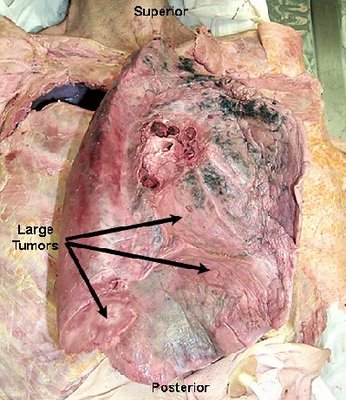
Doctors divide mesothelioma into different types based on what part of the mesothelium is affected. Mesothelioma most often affects the tissue that surrounds the lungs (pleura). This type is called pleural malignant mesothelioma. Other, rarer types of mesothelioma affect tissue in the abdomen, around the heart and around the testicles.

Mesothelioma is an aggressive and deadly form of cancer. Mesothelioma treatments are available, but for many people with mesothelioma, a cure is not possible. Instead, treatment for mesothelioma is often focused on keeping you as comfortable as possible.

Signs and symptoms of mesothelioma vary depending on where the cancer occurs.
Pleural mesothelioma, which affects the tissue that surrounds the lungs, causes signs and symptoms that may include; Chest pain under the rib cage, Painful coughing, Shortness of breath, Unusual lumps of tissue under the skin on your chest, Unexplained weight loss, Peritoneal mesothelioma, which occurs in tissue in the abdomen, causes signs and symptoms that may include:
Abdominal pain, Abdominal swelling, Lumps of tissue in the abdomen, Unexplained weight loss

Different forms of mesothelioma

Signs and symptoms of other types of mesothelioma are unclear. Other forms of mesothelioma are so rare that not much information is available. Pericardial mesothelioma, which affects tissue that surrounds the heart, can cause signs and symptoms such as breathing difficulty and chest pains. Mesothelioma of tunica vaginalis, which affects tissue surrounding the testicles, may be first detected as a mass on a testicle.

Check with your doctor

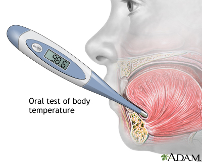
See your doctor if you have signs and symptoms that may indicate mesothelioma. Signs and symptoms of mesothelioma aren't specific to this disease and, due to the rarity of mesothelioma, are more likely to be related to other conditions. If any persistent signs and symptoms seem unusual or bothersome to you, ask your doctor to evaluate them.

Causes of Mesothelioma

In general, cancer begins when a series of genetic mutations occur within a cell, causing the cell to grow and multiply out of control. It isn't clear what causes the initial genetic mutations that lead to mesothelioma, though researchers have identified factors that may increase the risk. It's likely that cancers form because of an interaction between many factors, such as inherited conditions, your environment, your health conditions and your lifestyle choices.

Mesothelioma doesn't include a noncancerous tumor. A form of noncancerous (benign) tumor that can occur in the chest is sometimes called benign mesothelioma. However, this name is misleading. Benign mesothelioma doesn't begin in the same cells where the cancerous forms of mesothelioma begin. And, in a minority of cases, benign mesothelioma can be very aggressive, despite the term "benign." For this reason, some doctors now refer to this tumor as solitary fibrous tumor.

Solitary fibrous tumor usually doesn't cause signs and symptoms. Most cases are inadvertently discovered during tests and procedures for other conditions. It isn't clear what causes solitary fibrous tumors, but they aren't linked to asbestos exposure. Treatment for solitary fibrous tumor typically includes surgery.

Asbestos exposure: The primary risk factor for mesothelioma

Asbestos is a mineral that's found naturally in the environment. Asbestos fibers are strong and resistant to heat, making them useful in a wide variety of applications, such as in insulation, brakes, shingles, flooring and many other products.
When asbestos is broken up, such as during the mining process or when removing asbestos insulation, dust may be created. If the dust is inhaled or swallowed, the asbestos fibers will settle in the lungs or in the stomach, where they can cause irritation that may lead to mesothelioma, though how exactly this happens isn't understood. It can take 20 to 40 years or more for mesothelioma to develop as a result of asbestos exposure.
Some people with years of asbestos exposure never develop mesothelioma. And yet, others with very brief exposure develop the disease. This indicates that other factors may be involved in determining whether someone gets mesothelioma or doesn't. For instance, you could inherit a predisposition to cancer or some other condition could increase your risk.

Possible risk factors

Factors that may increase the risk of mesothelioma include:
Personal history of asbestos exposure. If you've been directly exposed to asbestos fibers at work or at home, your risk of mesothelioma is greatly increased.
Living with someone who works with asbestos. People who are exposed to asbestos may carry the fibers home on their skin and clothing. These stray fibers can put others in the home at risk of mesothelioma. People who work with asbestos should shower and change clothes before leaving work.

A monkey virus used in polio vaccines. Some research indicates a link between mesothelioma and simian virus 40 (SV40), a virus originally found in monkeys. Millions of people may have been exposed to SV40 when receiving polio vaccinations between 1955 and 1963, because the vaccine was developed using monkey cells. Once it was discovered that SV40 was linked to certain cancers, the virus was removed from the polio vaccine. Whether SV40 increases the risk of mesothelioma is a point of debate, and more research is needed.
Radiation. Some research links mesothelioma to the radioactive substance thorium dioxide, which was used along with X-rays to diagnose various health conditions from the 1920s to the 1950s. Mesothelioma has also been linked to radiation therapy treatments to the chest, such as those used to treat breast cancer or lymphoma.
Family history. A family history of mesothelioma may increase your risk of mesothelioma, but more research is needed to understand this theory.

Mesothelioma Complications
As pleural mesothelioma spreads in the chest, it puts pressure on the structures in that area. This can cause complications, such as:
  •     Difficulty breathing, Chest pain, Difficulty swallowing, 
  •     Swelling of the neck and face caused by pressure on the large vein that leads from your upper body to your heart (superior vena cava syndrome)
  •     Pain caused by pressure on the nerves and spinal cord, Accumulation of fluid in the chest (pleural effusion), which can compress the lung nearby and make breathing difficult

Prevention
Reducing your exposure to asbestos may lower your risk of mesothelioma.
Find out whether you work with asbestos environment
Most people with mesothelioma were exposed to the asbestos fibers at work. Workers who may encounter asbestos fibers include:
  •       Miners, Factory workers, Insulation manufacturers, Ship builders, Construction workers, Auto mechanics

Ask your employer whether you have a risk of asbestos exposure on the job.
Follow your employer's safety regulations
Follow all safety precautions in your workplace, such as wearing protective equipment. You may also be required to shower and change out of your work clothes before taking a lunch break or going home. Talk to your doctor about other precautions you can take to protect yourself from asbestos exposure.
Be safe around asbestos in your home
Older homes and buildings may contain asbestos. In many cases, it's more dangerous to remove the asbestos than it is to leave it intact. Breaking up asbestos may cause fibers to become airborne, where they can be inhaled. Consult experts trained to detect asbestos in your home. These experts may test the air in your home to determine whether the asbestos is a risk to your health. Don't attempt to remove asbestos from your home — hire a qualified expert. The Environmental Protection Agency offers advice on its website for dealing with asbestos in the home.