SAY NO TO DRUGS
Showing posts with label Nursing Science. Show all posts

Thursday, October 28, 2010

The Mental Status Exam

The mental status exam, is an assessment tool that helps identify psychological symptoms that may assist the practitioner determine if there is a psychogenic problem. When assessing mental status, it is important to adjust questions and categories to avoid age and/or cultural bias.
Category Description
Appearance General appearance, grooming and gait. This is best observed as the client comes into the room. Grooming is one of the earliest areas to deteriorate when mental function has diminished.
Behavior Speech, eye contact, body language, response to the environment. Observe for appropriate use of personal space. Does the person come right into your face, or stand an unusual distance away.
Insight The ability of the client to be aware of one’s own abilities. The ability to analyze a problem objectively. Ask the client to explain a problem.
Intellectual Functioning Simple calculations, ability to abstract or think symbolically and categories of association. This is done through direct questioning using math, proverbs or analogy.
Judgment Assesses decision-making abilities. Ask client What he would do in a dilemma regarding an important decision.
Memory Immediate recall, recent memory, remote memory. Ask the client about a recent current event that both you and the client should know. Ask about some event in the past that should be known by both. Be very careful in this area to avoid cultural bias.
Mood and Affect Mood relates to the emotions of the moment while affects refers to the range of emotions displayed such as happy, sad, or unchanging. Compare in relation the client’s probable everyday behavior.
Orientation Assess for awareness of person, time, place, and purpose.
Perceptual Processes Awareness of self and one’s thoughts, reality and fantasy. Ask about delusions, illusions and hallucinations. Do not hesitate do ask direct questions.
Sensorium Ability to concentrate, perception of stimuli.
Thought Contents This assesses themes in conversation and is assessed by observing what the client discusses spontaneously in conversation.
Thought Processes This measures a stream of conscious or mental activity as indicated in speech. Observe for rate, flow, and ability to pursue a topic logically.


www.accessce.com

Neurological Assessment : Checks Pupils

Neurological Assessment : Checks Pupils


Neurological Assessment : Checks Pupils


  1. Observes Both Pupils Simultaneously For: Equality, Size and Shape.
    • Compares pupils for equality.
    • Determines size, dilated, constricted, pinpoint.
    • Determines irregularities in shape.

  2. Observes Direct Pupillary Light Reflexes.
    • Checks one pupil at a time.
    • Shines flashlight into eye from side.
    • Repeat other eye.

  3. Observes Consensual Pupillary Reflex
    • Shines flashlight into each eye alternately.
    • Observes opposite pupil. Opposite pupil should constrict when light shore.
    • Charts description of pupils: Equality, size, shape, reaction to light.

  4. Observes pupillary response to accommodation
    • Have patient follow a closer moving object such as a pen.
    • Pupils will constrict (or accommodate) to the closer moving object. * cannot be tested on blind or confused persons.

  5. Observes Extraocular Movements
    • Asks patient to focus on object.
    • Moves object; medical, lateral, superior, inferior and circular. In the pattern of an "H."
    • Observes movement of both eyes in each of above directions; notes abnormalities or weakness. A. Charts extraocular movements as "full" if no abnormalities or "unable to move eyes laterally, medially etc."
accessce.com

Wednesday, October 27, 2010

Vital Signs - Blood Pressure

Blood Pressure

Vital Signs - Blood Pressure


Blood Pressure : Pressure of blood against the walls of the arteries

Systolic : Number that is on the top, and when heart is contracting

Diastolic : Number that is on the bottom, and when heart is at rest

Systolic range : 90 - 140

Diastolic range : 60 - 90

Hypertension : High blood pressure, above 140 systolic or over 90 diastolic

Hypotension : Low blood pressure, under 90 over 60

To measure systolic : Sound of first beat

To measure diastolic : No beat is heard

Hypertension thickens heart muscle (hypertrophy) and reduces chamber in size

Thigh cuff for large arms, Small cuff pediatrics

Sphygmomanometer is instrument use to take blood pressure

Pulse pressure: Difference between systolic and diastolic

Vital Signs - Pulse

Pulse

Vital Signs - Pulse


Rate is : Number of beats per minute

Rhythm is : Regularity of beats

Normal range of adults : 60 - 100 per minute

Pulse : Can be weak, bounding or absent for short period of time

Rhythm : Can be regular or irregular

Palpate for : Rhythm, rate, and strength

Optimal finding : 80 per min. strong, and reg.

Tachycardia : Over 100 beats per minute

Bradycardia : Under 60 beats per minute

To measure pulse : count 30 sec.X 2

For irregular pulse : count the full 60seconds

Auscultate : Use stethoscope

Pulse deficit : Difference of apical and radial.

Vital Signs Respirations

Respirations


Vital Signs Respirations


Respirations : How many breaths per minute

Adults: 12 - 20 / Infant slightly higher 20 - 40

Inhalation and Exhalation equals: 1 breath

To count breaths: Count 30 seconds by 2

Look for : Rhythm, rate, depth, and quality

Bradypnea: Under 12 breaths

Tachypnea: Over 20 breaths

Eupnea: Normal rate and depth

Apnea: Not breathing maybe 30 seconds or at all

Dyspnea: Difficulty in breathing

Orthopnea: Over bedside 90o postural position

Hyperpnea: Fast respirations

Cheyne Stokes: Increasing in rate and depth then periods of apnea - cyclic.

Kussmaul: Metabolic acidosis,usually the Diabetic. Rapid, very deep respirations intended to blow off carbondioxide.

Vital Signs - Temperature

Temperature


Vital Signs - Temperature


Oral - mouth

Time period 3 minutes
Normal range: 97.6 - 99.6 degrees
Absolute: 98.6 degrees

Rectal - Anus
Time period 3 minutes
Position -Lateral Sims
Normal range: 98.6 - 100.6 degrees
Absolute: 99.6 degrees

Axillary - Armpit
Time period 10 minutes
Normal range: 96.6 - 98.6
Absolute: 97.6 degrees
Otic or Tympanic Time period 10 sec. or less

Degree range is calibrated to rectal or oral
Hypothermia - Low body temperature
Hyperthermia - High body temperature
Pyrexia - High fever
Febrile - High fever
Afebrile - No fever

Things that can effect temperature: smoking, fluids, oxygen use, food, colds, or flu.(www.accessce.com)

Apgar Score

Apgar Scoring


Sign
0 points
1 point
2 points
A
Activity
(Muscle tone)
limp
limbs flexed
active movement
P
Pulse
(heart rate)
absent
<>
> 100 /min
G
Grimace
(response to smell or foot slap)
absent
grimace
cough or sneeze (nose)
cry and withdrawal of foot (foot slap)
A
Appearance
(color)
blue
body pink
extremities blue
black over
R
Respiration
(breathing)
absent
irregular
weak crying
good strong cry

The total Apgar score is the sum of the scores for the five signs.


The 12 Cranial Nerves

The 12 Cranial Nerves


There are 12 pairs of cranial nerves. These nerves arise from the brain and brain stem, carrying motor and or sensory information.


Cranial nerve I : Olfactory nerve

The olfactory nerve is composed of axons from the olfactory receptors in the nasal sensory epithelium. It carries olfactory information (sense of smell) to the olfactory bulb of the brain. This is a pure sensory nerve fiber.

Cranial nerve II: Optic nerve

The optic nerve is composed of axons of the ganglion cells in the eye. It carries visual information to the brain. This is a pure sensory nerve fiber. This nerve travels posteromedially from the eye, exiting the orbit at the optic canal in the lesser wing of the sphenoid bone. The optic nerves join each other in the middle cranial fossa to form the optic chiasm.


Cranial nerve III: Oculomotor nerve

The oculomotor nerve is composed of motor axons coming from the oculomotor nucleus and the edinger-westphal nucleus in the rostral midbrain located at the superior colliculus level. This is a pure motor nerve. It provides somatic motor innervation to four of the extrinsic eye muscles: the superior rectus, inferior rectus, medial rectus, and the inferior oblique muscles. It also innervates the muscles of the upper eyelid and the intrinsic eye muscles (the pupillary eye muscle.) Together, CN III, CN IV and CN VI control the six muscles of the eye.


Cranial nerve IV: Trochlear nerve

The trochlear nerve provides somatic motor innervation to the superior oblique eye muscle. This cranial nerve originates at the trochlear nucleus located in the tegmentum of the midbrain at the inferior colliculus level and exits the posterior side of the brainstem. It is also a pure motor nerve fiber.


Cranial nerve V: Trigeminal nerve

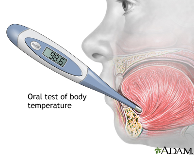
The trigeminal is the largest cranial nerve . It provides sensory information from the face, forehead, nasal cavity, tongue, gums and teeth (touch, and temperature) and provides somatic motor innervation to the muscles of mastication or “chewing”.

This cranial nerve has 3 branches: the ophthalmic, maxillary and mandibular branches.

It is composed of both sensory and motor axons. The sensory fibers are located in the trigeminal ganglion and the motor fibers project from nuclei in the pons.


Cranial nerve VI: Abducens nerve

The abducens nerve carries somatic motor innervation to one of the extrinsic eye muscles, the lateral rectus muscle. It is another pure motor nerve fiber and originates from the abducens nucleus located in the caudal pons at the facial colliculus level.


Cranial nerve VII: Facial nerve

The facial nerve carries somatic motor innervation to the many muscles for facial expression. It carries sensory information form the face (deep pressure sensation) and taste information from the anterior two thirds of the tongue. It arises at the pons in the brainstem and it emerges through openings in the temporal bone and stylomastoid foramen and has many branches. It is composed of both sensory and motor axons.


Cranial nerve VIII: Vestibulocochlear nerve

The vestibulocochlear nerve innervates the hair cell receptors of the inner ear. It carries vestibular information to the brain from the semicircular canals, utricle, and saccule providing the sense of balance. It also carries information from the cochlea providing the sense of hearing. This cranial nerve branches into the Vestibular branch (balance) and the cochlear branch (hearing). The cochlear fibers originate from the spiral ganglion. It is pure sensory nerve fiber.


Cranial nerve IX: Glossopharyngeal nerve

The glossopharyngeal nerve innervates the pharynx (upper part of the throat), the soft palate and the posterior one-third of the tongue. It carries sensory information (touch, temperature, and pressure) from the pharynx and soft palate. It carries taste sensation from the taste buds on the posterior one third of the tongue. It provides somatic motor innervation to the throat muscles involved in swallowing. It provides visceral motor innervation to the salivary glands. This cranial nerve also supplies the carotid sinus and reflex control to the heart . It is composed of both sensory and motor axons and originates from the nucleus ambiguous in the reticular formation of the medulla.


Cranial nerve X: Vagus nerve

The vagus nerve consists of many rootlets that come off of the brainstem just behind the glossopharyngeal nerve. The branchial motor component originates from the nucleus ambiguous in the reticular formation of the medulla. The visceral component originates from the dorsal motor nucleus of the vagus located in the floor of the fourth ventricle in the rostral medulla and in the central grey matt er of the caudal medulla. It is the longest cranial nerve
innervating many structures in the throat, including the muscles of the vocal cords, thorax and abdominal cavity. It provides sensory information (touch, temperature and pressure) from the external auditory meatus (ear canal) and a portion of the external ear. It carries taste sensation from taste buds in the pharynx. It also provides sensory information from the esophagus, respiratory tract, and abdominal viscera (stomach, intestines, liver, etc.). It provides visceral motor innervation to the heart, stomach, intestines, and gallbladder. It is part of the ANS, the parasympathetic branch. It is composed of both sensory and motor axons. Other parasympathetic ganglia include CN III , CN VII and CN IX .


Cranial nerve XI: Spinal Accessory nerve

The spinal accessory nerve has two branches. The cranial branch provides somatic motor innervation to some of the muscles in the throat involved in swallowing. This cranial branch is accessory to CN X, originating in the caudal nucleus ambiguous, with the fibers of the cranial root traveling the same extracranial path as the branchial motor component of the vagus nerve. The spinal branch provides somatic motor innervation to the trapezius muscles, providing muscle movement for the upper shoulders head and neck. It is pure motor nerve fiber.


Cranial nerve XII: Hypoglossal nerve

The hypoglossal nerve provides somatic motor innervation to the muscles of the tongue. This pure motor nerve originates from the hypoglossal nucleus located in the tegmentum of the medulla.


Source : www.pitt.edu

Normal Heart Sounds

Normal Heart Sounds

Normal Heart Sounds

Heart Sounds

The heart sounds are the noises (sound) generated by the beating heart and the resultant flow of blood through it. This is also called a heartbeat. In cardiac auscultation, an examiner uses a stethoscope to listen for these sounds, which provide important information about the condition of the heart.

In healthy adults, there are two normal heart sounds often described as a lub and a dub (or dup), that occur in sequence with each heart beat. These are the first heart sound (S1) and second heart sound (S2), produced by the closing of the AV valves and semilunar valves respectively. In addition to these normal sounds, a variety of other sounds may be present including heart murmurs, adventitious sounds, and gallop rhythms S3 and S4.

Heart murmurs are generated by turbulent flow of blood, which may occur inside or outside the heart. Murmurs may be physiological (benign) or pathological (abnormal). Abnormal murmurs can be caused by stenosis restricting the opening of a heart valve, resulting in turbulence as blood flows through it. Abnormal murmurs may also occur with valvular insufficiency (or regurgitation), which allows backflow of blood when the incompetent valve closes with only partial effectiveness. Different murmurs are audible in different parts of the cardiac cycle, depending on the cause of the murmur.


Normal Heart Sounds

Normal heart sounds are associated with heart valves closing, causing changes in blood flow.

S1

The first heart tone, or S1, forms the "lubb" of "lubb-dub" or "lubb-dup" and is composed of components M1 and T1. Normally M1 precedes T1 slightly. It is caused by the sudden block of reverse blood flow due to closure of the atrioventricular valves, i.e. mitral and tricuspid, at the beginning of ventricular contraction, or systole. When the ventricles begin to contract, so do the papillary muscles in each ventricle. The papillary muscles are attached to the tricuspid and mitral valves via chordae tendineae, which bring the cusps or leaflets of the valve closed (chordae tendineae also prevent the valves from blowing into the atria as ventricular pressure rises due to contraction). The closing of the inlet valves prevents regurgitation of blood from the ventricles back into the atria. The S1 sound results from reverberation within the blood associated with the sudden block of flow reversal by the valves.[1] If T1 occurs more than slightly after M1, then the patient likely has a dysfunction of conduction of the right side of the heart such as a Right bundle branch block.

S2

The second heart tone, or S2, forms the "dub" of "lubb-dub" or "lubb- dup" and is composed of components A2 and P2. Normally A2 precedes P2 especially during inspiration when a split of S2 can be heard. It is caused by the sudden block of reversing blood flow due to closure of the aortic valve and pulmonary valve at the end of ventricular systole, i.e. beginning of ventricular diastole. As the left ventricle empties, its pressure falls below the pressure in the aorta, aortic blood flow quickly reverses back toward the left ventricle, catching the aortic valve pocketlike cusps and is stopped by aortic (outlet) valve closure. Similarly, as the pressure in the right ventricle falls below the pressure in the pulmonary artery, the pulmonary (outlet) valve closes. The S2 sound results from reverberation within the blood associated with the sudden block of flow reversal.

Splitting of S2 normally occurs during inspiration because the decrease in intrathoracic pressure causes more blood to be delivered to the right heart, thereby prolonging contraction and delaying closure of the pulmonic valve. A widely split S2 can be associated with several different cardiovascular conditions, including right bundle branch block and pulmonary stenosis.(wikipedia)

Assessing Lung Sound

Assessing Lung Sound


Assessing Lung Sound

To auscultate lung sounds, move the diaphragm of your stethoscope according to the numbers on the corresponding diagram.

There are three normal breath sounds :
  • (B) Bronchial Breath Sounds - loud, harsh, hight pitched
    Heard over trachea, bronchi (betwen clavicles and midsternum), and over main bronchus.

  • (BV) Bronchovesicular Breath Sounds - blowing sounds, moderate intensity and pitch.
    Heard over large airways, on either side of sternum, at the Angle of Louis, and betwen scapulae.

  • (V) Vesicular Breath Sounds - soft breezy quality, low pitched.
    Heard over the peripheral lung area, heard at best of base lungs.

Tuesday, October 26, 2010

Glasgow Coma Scale

Glasgow Coma Scale

The Glasgow Coma Scale provides a score in the range 3-15; patients with scores of 3-8 are usually said to be in a coma. The total score is the sum of the scores in three categories. For adults the scores are as follows:

Eye Opening Response Spontaneous--open with blinking at baseline 4 points
Opens to verbal command, speech, or shout 3 points
Opens to pain, not applied to face 2 points
None 1 point
Verbal Response Oriented 5 points
Confused conversation, but able to answer questions 4 points
Inappropriate responses, words discernible 3 points
Incomprehensible speech 2 points
None 1 point
Motor Response Obeys commands for movement 6 points
Purposeful movement to painful stimulus 5 points
Withdraws from pain points
Abnormal (spastic) flexion, decorticate posture 3 points
Extensor (rigid) response, decerebrate posture 2 points
None 1 point


Source : www.unc.edu