|
|---|
Friday, April 30, 2010
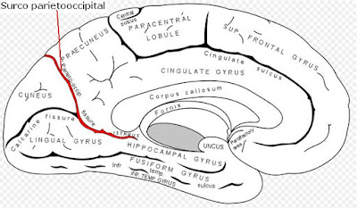
Surco parietooccipital

Thursday, April 29, 2010
Cisura Calcarina

Wednesday, April 28, 2010
Corteza entorrinal

Tuesday, April 27, 2010
Circunvolución angular
La circunvolución angular del cerebro humano se encuentra de tres a cuatro veces más desarrollada que en el resto de los primates, y está ubicada estratégicamente en una área adyacente, o contígua, al área de procesamientos de información táctil, de la comprensión del lenguaje escrito y hablado (área de Wernicke), y de la visión. De acuerdo a las últimas investigaciones realizadas por el Dr V. S. Ramachandran de la Universidad de California, la circunvolución angular participaría en los procesos corticales de la comprensión de las metáforas conceptuales y otras abstracciones.
Localización de la circunvolución angular
Monday, April 26, 2010
Circunvolución supramarginal

Saturday, April 24, 2010
Circunvolución parietal inferior
La circunvolución parietal inferior comprende las áreas de Brodmann 39 y 40 y está dividida en dos circunvoluciones: la supramarginal o circunfleja, la cual se arquea alrededor del extremo de la cisura de Silvio, y la angular. Sus funciones son: 1) Esterognosia, que es la capacidad de reconocer los objetos por el tacto y su falta se denomina Asterognosia. 2) Lexia, la cual es la capacidad de leer y comprender lo que se lee; su lesión cuasa alexia. 3) calculia: capacidad de realizar cálculos matemáticos sencillos.

Friday, April 23, 2010
Development of Nursing Theories
Basic processes in the development of nursing theories
Nursing theories are often based on & influenced by broadly applicable processes & theories. Following theories are basic to many nursing concepts.
General System Theory
It describes how to break whole things into parts & then to learn how the parts work together in “systems”. These concepts may be applied to different kinds of systems, e.g. Molecules in chemistry, cultures in sociology, and organs in Anatomy & Health in Nursing.
Adaptation Theory
It defines adaptation as the adjustment of living matter to other living things & to environmental conditions.
Adaptation is a continuously occurring process that effects change & involves interaction & response.
Human adaptation occurs on three levels :
- 1. The internal (self)
- 2. The social (others) &
- 3. the physical (biochemical reactions)
Developmental Theory
- It outlines the process of growth & development of humans as orderly & predictable, beginning with conception & ending with death.
- The progress & behaviors of an individual within each stage are unique.
- The growth & development of an individual are influenced by heredity, temperament, emotional, & physical environment, life experiences & health status.
Common concepts in nursing theories
Four concepts common in nursing theory that influence & determine nursing practice are:
- The person (patient).
- The environment
- Health
- Nursing (goals, roles, functions)
Each of these concepts is usually defined & described by a nursing theorist, often uniquely; although these concepts are common to all nursing theories. Of the four concepts, the most important is that of the person. The focus of nursing, regardless of definition or theory, is the person.
Thursday, April 22, 2010
Circunvolución parietal superior

Nursing Care Plan for Pneumonia

Nursing Plan
Breath Pattern ineffectiveness because of pulmonary infection
Characteristics :
Cough (both productive and non productive), shortness of breath, Tachipnea, breath sounds are limited, retraction, fever, diaporesis, ronchii, cyanosis, leukocytosis.
Goal :
Effective breathing pattern characterized by :
- Voice of lung breath clean and the same on both sides
- The temperature of the body within the limits of 36.5 to 37.2 OC
- The rate of breathing in the normal range
- There is no coughing, cyanosis, retraction and diaporesis
Intervention :
- Perform assessments every 4 hours of respiratory rate, temperature, and signs of airway effectiveness.
Rational: Evaluation and reassessment of the actions that will be / have been granted. - Perform scheduled Phisioterapi chest
Rational: Removing the secretion of the airway, preventing obstruction - Give Oxygen
Rational: Increased lung tissue oxygen supply - Give antibiotics and antipyretics, assess the effectiveness and side effects (rash, diarrhea)
Rational: Eradication of the bacteria as a factor of disturbance causa - Make checks thoracic photo
Rational: The evaluation of the effectiveness of the circulation of oxygen, evaluated the condition of lung tissue - Perform a gradual suction
Rational: Helping airway clearance - Record the results of the pulse oximeter when installed, every 2 - 4 hours
Rational: Periodically Evaluate the success of therapy / health team action.
Wednesday, April 21, 2010
Home Care : Breast Pain
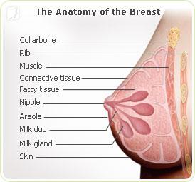
Home Care
For tips on how to manage pain from fibrocystic breasts, see breast lumps.
Certain birth control pills may help relieve breast pain. Ask your doctor if this therapy is right for you.
If you have a breast infection, you may need antibiotics. Look for symptoms of infection such as redness in the area, nipple discharge, or fever. Contact your doctor if you have these symptoms.
If you have a breast injury, immediately apply a cold compress such as an ice pack (wrapped in a cloth -- don't apply directly to the skin) for 15 to 20 minutes. Take a nonsteroidal anti-inflammatory drug (NSAID) such as ibuprofen to reduce your chance of developing persistent breast pain or swelling.
http://ncp-nursingcareplans.blogspot.com
www.nlm.nih.gov
Circunvolución parietal ascendente

Tuesday, April 20, 2010
Home Care : Nausea and Vomiting
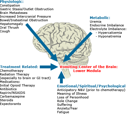
Home Care
It is important to stay hydrated. Try frequent, small amounts of clear liquids, such as electrolyte solutions. Other clear liquids -- such as water, ginger ale, or fruit juices -- also work unless the vomiting is severe or it is a baby who is vomiting.
For breast-fed babies, breast milk is usually best. Formula-fed babies usually need clear liquids.
Don't drink too much at one time. Stretching the stomach can make nausea and vomiting worse. Avoid solid foods until there has been no vomiting for six hours, and then work slowly back to a normal diet.
An over-the-counter bismuth stomach remedy like Pepto-Bismol is effective for upset stomach, nausea, indigestion, and diarrhea. Because it contains aspirin-like salicylates, it should NOT be used in children or teenagers who might have (or recently had) chickenpox or the flu.
Most vomiting comes from mild viral or food-related illnesses. Nevertheless, if you suspect the vomiting is from something serious, the person may need to be seen immediately by a medical professional.
If you have morning sickness during pregnancy, ask your doctor about the many possible treatments.
The following may help treat motion sickness:
* Lying down
* Over-the-counter antihistamines (such as Dramamine)
* Scopolamine prescription skin patches (such as Transderm Scop) are useful for extended trips, such as an ocean voyage. Place the patch 4 - 12 hours before setting sail. Scopolamine is effective but may produce dry mouth, blurred vision, and some drowsiness. Scopolamine is for adults only. It should NOT be given to children.
www.nlm.nih.gov
Circunvolución cingulada

Corte transversal del cerebro que muestra la circunvolución cingulada

Monday, April 19, 2010
Home Care : Diarrhea

Home Care
- Drink plenty of fluid to avoid becoming dehydrated. Start with sips of any fluid other than caffeinated beverages. Milk may prolong loose stools, but also provides needed fluids and nourishment. Drinking milk may be fine for mild diarrhea. For moderate and severe diarrhea, electrolyte solutions available in drugstores are usually best.
- Active cultures of beneficial bacteria (probiotics) make diarrhea less severe and shorten its duration. Probiotics can be found in yogurt with active or live cultures and in supplements.
- Foods like rice, dry toast, and bananas can sometimes help with diarrhea.
- Avoid over-the-counter antidiarrhea medications unless specifically instructed to use one by your doctor. Certain infections can be made worse by these drugs. When you have diarrhea, your body is trying to get rid of whatever food, virus, or other bug is causing it. The medicine interferes with this process.
- Get plenty of rest.
If you have a chronic form of diarrhea, like the one caused by irritable bowel syndrome, try adding bulk to your diet -- to thicken the stool and regulate bowel movements. Such foods include fiber from whole-wheat grains and bran. Psyllium-containing products such as Metamucil or similar products can also add bulk to stools.
www.nlm.nih.gov
Cisura calloso marginal
Ubicación anatómica de la cisura calloso marginal en cara interna del hemisferio cerebral izquierdo

Sunday, April 18, 2010
Fascículo longitudinal superior

Saturday, April 17, 2010
Debridement
One way is to use a scalpel and special scissors.
- The skin surrounding the wound is cleaned and disinfected.
- The wound is probed with a metal instrument to determine how deep it is and to see if there is any foreign material or object in the ulcer.
- The doctor cuts away the dead tissue, then washes out any unattached tissue.
- Your sore may seem bigger and deeper after the doctor or nurse debrides it. The ulcer should be red or pink in color and look like fresh meat.
Other ways to remove dead or infected tissue are to :
- Put your foot in a whirlpool bath.
- Use a syringe and catheter (tube).
- Apply wet to dry dressings to the area.
- Put special chemicals, called enzymes, on your ulcer. These dissolve dead tissue from the wound.
Células de Martinotti

Friday, April 16, 2010
Diabetes - taking care of your feet
Diabetes can damage the nerves and blood vessels in your feet. This damage can cause numbness and reduce feeling in your feet. As a result, your feet may not heal well if they are injured. If you get a blister, you may not notice, and it may get worse.
Check your feet every day. Inspect the top, sides, soles, heels, and between the toes. Look for:
- Dry and cracked skin
- Blisters or sores
- Bruises or cuts
- Redness, warmth, or tenderness
- Firm or hard spots
If you cannot see well, ask someone else to check your feet.
Call your doctor right way about any foot problems. Do not try to treat them yourself first. Even small sores or blisters can become big problems if infection develops or they do not heal.
Wash your feet every day with lukewarm water and mild soap. Strong soaps may damage the skin.
- Check the temperature of the water with your hands or elbow first.
- Gently dry your feet, especially between the toes.
- Use lotion, petroleum jelly, lanolin, or oil on dry skin. Do NOT put lotion between your toes.
Ask your health care provider to show you how to trim your toenails.
- Soak your feet in lukewarm water to soften the nail before trimming.
- Cut the nail straight across, because curved nails are more likely to become ingrown.
- Your foot doctor (podiatrist) can trim your nails if you are unable to.
Most people with diabetes should have corns or calluses treated by a foot doctor. If your doctor has given you permission to treat corns or calluses on your own:
- Gently use a pumice stone to remove corns and calluses after a shower or bath, when your skin is soft.
- Do NOT use medicated pads or try to shave or cut them away at home.
If you smoke, stop. Smoking decreases blood flow to your feet. Talk with your doctor or nurse if you need help quitting.
Do not use a heating pad or hot water bottle on your feet. Do not walk barefoot, especially on hot pavement or hot sandy beaches. Remove your shoes and socks during visits to your health care provider so that they can check your feet.
Source : http://www.nlm.nih.gov/medlineplus/ency/patientinstructions/000081.htm
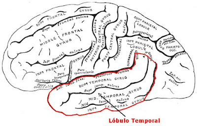
Lóbulo temporal
Thursday, April 15, 2010
Circunvolución temporal inferior

Wednesday, April 14, 2010
Capas de la corteza cerebral
1) Capa molecular, o plexiforme: es la más superficial. Consiste en una red densa de fibras nerviosas orientadas tangencialmente. Estas derivan de dendritas de células piramidales y fusiformes, los axones de células estrelladas y de Martinotti. También hay fibras aferentes que se originan en el tálamo, de asociación y comisurales. Entre las fibras nerviosas hay algunas células de Cajal. Por ser la capa más superficial se establecen gran cantidad de sinapsis enter diferentes neuronas.
2) Capa granular externa: contiene un gran número de pequeñas células piramidales y estrelladas. Las dendritas de éstas células terminan en la capa molecular y los axones entran en las capas más profundas.
3) Capa piramidal externa: esta capa está compuesta por células piramidales. Su tamaño aumenta desde el límite superficial hasta el límite más profundo. Las dendritas pasan hasta la capa molecular y los axones hasta la sustancia blanca como fibras de proyección, asociación o comisurales.
4) Capa granular interna: esta capa está compuesta por células estrelladas dispuestas en forma muy compacta. Hay una gran concentración de fibras dispuestas horizontalmente conocidas en conjunto como la banda externa de Baillarger.
5) Capa ganglionar (capa piramidal interna): esta capa contiene células piramidales muy grandes y de tamaño mediano. Entre las células piramidales hay células estrelladas y de Martinotti. Además hay un gran número de fibras dispuestas horizontalmente que forman la banda interna de Baillger. En las zonas motoras de la circunvolución precentral, las células de proyección de Betz dan origen aproximadamente al 3% de las fibras de proyección del haz corticoespinal.
6) Capa multiforme (capa de células polimórficas): aunque la mayoría de las células son fusiformes, muchas son células piramidales modificadas cuyo cuerpo celular es triangular u ovoideo. Las células de Martinotti también son conspicuas en esta capa. Hay muchas fibras nerviosas que entran en la sustancia blanca subyacente.
Tuesday, April 13, 2010
Picture Your Heart's Health With EKGs
by: Damian Sofsian
Each time your heart beats, the contractions and relaxations of the heart muscle emit electrical current. An electrocardiogram (EKG) is a medical recording of the electric impulses from the heart. Electrodes that send impulses to the EKG machine are attached to the patient�s skin at various points on the body. Those recorded currents are displayed on a computer monitor and can be printed out on special graph paper. Your heart�s electrical currents are recorded on the graph paper as an EKG. Qualified medical staff interpret the graphed results to determine any irregularities.
Most EKGs are performed in a critical care facility, telemetry or any place that a particular patient needs to be monitored. EKGs can help your doctor determine the status of your heart health. By graphing the electrical impulses of the heart, doctors and other trained medical staff are able to see the presence of any abnormalities. The EKG recording often reveals the scars of past heart attacks and other heart damage. Although the test cannot predict future heart attacks or other heart problems, a combination of family history and additional examinations may give your doctor a good idea of what to expect.
Individuals experiencing chest pain, shortness of breath, dizziness or heart palpitations will likely be referred for an EKG by their doctor. An EKG is a rapid and safe way to determine if a heart attack is occurring. Those reporting these types of symptoms will likely be referred to the nearest Emergency Room for further evaluation. If your doctor does not think your symptoms indicate a life-threatening situation, you may be asked to make an appointment with an EKG specialist for further observation.
An EKG is a very simple and painless procedure. The patients are instructed to lie face up on an examination table while electrodes are strategically placed at various points on their body. The electrodes are attached to cables and the cables are attached to the EKG machine. The electrodes send electronic impulses to the machine and results in a printed graph, which is a picture of your heart function. The procedure usually takes 15 to 20 minutes but may require a longer visit if the technician needs additional testing data. A stress test is a normal EKG procedure that requires the patient perform moderate exercise while recording heart rhythms.
EKG Reading provides detailed information on EKG, EKG Reading, EKG Interpretation, EKG Machine and more. EKG Reading is affiliated with ECG Cross Reference.
To find other free health content see e-healtharticles.com
http://askep-asuhankeperawatan.blogspot.com/2010/04/picture-your-hearts-health-with-ekgs.html
Circunvolución temporal media

Monday, April 12, 2010
El caso de Phineas Gage


Saturday, April 10, 2010
Nursing Care Plan For Myocardial Infarction
Myocardial infarction (MI) is the rapid development of myocardial necrosis caused by a critical imbalance between oxygen supply and demand of the myocardium. This usually results from plaque rupture with thrombus formation in a coronary vessel, resulting in an acute reduction of blood supply to a portion of the myocardium.
Possible causes of Myocardial infarction (MI) are : Coronary artery occlusion, Coronary spasm and Coronary stenosis. There are some risk factors to develop of Myocardial infarction such as :
- Aging
- Decrease serum HDL levels
- Diabetes Mellitus
- Drug use, specifically use of amphetamines or cocaine
- Elevated serum Triglyceride, LDL and Cholesterol levels
- Excessive intake of saturated fats, carbohydrates, or salt
- Family history of CAD
- Hypertension
- Obesity
- Post menopausal women
- Sedentary lifestyle
- Smoking
- Stress
Nursing Care Plan For Myocardial Infarction :
Assessment findings on the patient with myocardial infarction are : Dyspnea, Diaphoresis, Arrhythmias, Tachicardia, Anxiety, Pallor, Hypotension, Nausea and vomiting, Elevated temperature. The specific complain from the patient is crushing substernal chest pain (may radiate to the jaw, back and arms) that unrelieved by rest or nitroglycerin (NGT) tablet.
Nursing Diagnoses:
- Chest discomfort (pain) due to an inbalance Oxygen (O2) demand supply
- Potential Arrhythmias related to decrease cardiac output
- Respiratory difficulties (dyspnoea) due to decrease CO
- Anxiety & fear of death related to his condition
- Activity intolerance related to limitations imposed
- Potential for complications of thrombolytic therapy
- Discharge medications, follow up & Health teachings
Planing and goals :
- The patient won't develop preventable complication
- The patient will understand the necessary treatment and lifestyle changes.
Intervention:
- Monitor ECG result to detect ischemia, injury new or extended infarction, arrhythmia, and conduction defects
- Monitor, record vital signs and hemodynamic variables to monitor response to the therapy and detects complication
- Administer oxygen as prescribe to improve oxygen supply to the heart
- Obtain an ECG reading during acute pain to detect myocardial ischemia, injury or infarction
- Maintain the patient's prescribed diet to reduce fluid retention and cholesterol levels
- Provided postoperative care if necessary to avoid postoperative complications and help the patient achieve a full recovery
- Allay the patient's anxiety because the anxiety increase oxygen demands.
Corteza auditiva primaria
Las neuronas de la corteza auditiva primaria están organizadas de acuerdo a las frecuencias de los sonidos a las cuales ellas responden mejor. La neuronas el extremo anterior de la corteza auditiva responden mejor a las bajas frecuencias, mientras que las otras ubicadas en el extremo posterior responden mejor a las frecuencias altas. Una lesión unilateral produce sordera parcial en ambos oídos con mayor pérdida del lado contralateral.
La corteza auditiva secundaria está ubicada detrás del área auditiva primaria. Se cree que esta área es necesaria para la interpretación de los sonidos.
 Corteza auditiva primaria: 41 - 42
Circunvoluciones temporales transversas

Friday, April 9, 2010
WHO : Experts begin their assessment of the response to the H1N1 influenza pandemic
Dr Margaret Chan
Director-General of the World Health Organization
Excellencies, distinguished members of the Review Committee, representatives of member states, colleagues in the UN system, representatives of nongovernmental organizations, members of the media, ladies and gentlemen,
I am pleased to welcome you to the start of this review process. I am also pleased to see such a broad range of interests and expertise represented in this room.
This has been the first influenza pandemic in four decades. This has been the first major test of the functioning of the revised International Health Regulations, which entered into force in 2007.
The International Health Regulations have a provision that calls for a review of their functioning no later than five years after their entry into force. In 2008, the World Health Assembly decided that this first review should be undertaken by the Sixty-third World Health Assembly in May 2010.
As you know, this provision and this decision were in place prior to the onset of the 2009 H1N1 influenza pandemic.
During the January 2010 session of the Executive Board, I proposed that the scheduled IHR review could also be used to assess the international response to the influenza pandemic. The Executive Board agreed to this proposal.
I believe there is merit in assessing the performance of an international instrument, like the IHR, when put to an extreme test by a widespread and closely scrutinized infectious disease event.
As I have said before, this has been the most closely watched and carefully scrutinized pandemic in history. This gives us a vast body of scientific, clinical, and epidemiological data to assess.
Moreover, the pandemic’s spread was rapidly global. To date, laboratory confirmed cases of H1N1 pandemic influenza have been officially reported from 213 countries and overseas territories or communities. This gives us a vast and varied experience to assess.
The outbreak of SARS, the first severe new disease of the 21st century, occurred in 2003 while drafting of the revised Regulations was under way. Experiences during that outbreak led to many refinements in the Regulations, including the introduction of a system of checks and balances to ensure that no one, myself included, has unfettered power.
I see potential advantages in assessing the performance of the Regulations with a particular focus on the influenza pandemic and how it was managed, especially at the international level by WHO. When the performance of the IHR is assessed under the challenging conditions of an influenza pandemic, specific strengths and weaknesses are likely to come to light.
Ladies and gentlemen,
In planning and organizing this review, WHO aimed to facilitate a process that is independent, credible, and transparent. We want a frank and critical assessment. WHO is not defining or restricting the scope of specific issues that may arise. If our member states have questions or concerns, we want to hear these questions and concerns raised.
We are seeking lessons, about how the IHR has functioned, about how WHO and the international community responded to the pandemic, that can aid the management of future public health emergencies of international concern. And I can assure you: there will be more.
We want to know what worked well. We want to know what went wrong and, ideally, why. We want to know what can be done better and, ideally, how.
In a spirit of inclusiveness, this meeting has been opened to a range of organizations and agencies interested in improving our collective management of public health emergencies. We want to hear your views as well.
To support the credibility and independent nature of the review process, the Secretariat has been diligent in inviting a membership in this committee that is geographically balanced, that includes the views and experiences of developing and developed countries, and that represents a broad range of scientific expertise and practical experience in multiple disciplines.
The Secretariat has also been especially vigilant in seeking out possible conflicts of interest among committee members.
As I said, we want a frank, critical, transparent, credible and independent review of our performance, as well as that of the International Health Regulations. The Secretariat will do everything it can to facilitate such a process.
Thank you.
Source : http://www.who.int
Fascículo arcuato


Thursday, April 8, 2010
Circunvolución temporal superior

Nursing Care Plan For Acute Renal Failure
Implement intervention to prevent infection and the complications of immobility. Because She/He is on bed rest, the client becomes susceptible to the hazards of immobility. Infection is a serious risk and the leading cause of death in client with acute renal failure.
Assessment
During assessment, the nurses may find some sign and symptom of acute renal failure. There are many complain from patient related to his/her condition such as ; Anorexia, Nausea, Vomiting, Costovertebral plain, Headache, diarrhea or constipation, Irritability, Restlessness, Lethargy, Drowsiness, Stupor, Coma, Pallor, Ecchymosis, Stomatitis, Thick tenaciouse sputum, Urine output less than 400 ml/day for 1 to 2 weeks and then followed by diuresis (3 to 5 L/day) for 2 to 3 weeks, Weight gain.
Nursing Diagnosis
- Ineffective tissue perfusion (renal)
- Excess fluid volume
- Risk for infection
- Risk for deficient fluid volume.
Planing and Goal
- The client will have normal fluid and electrolyte levels
- The client will experience no preventable complication
- The client will understand the means by which His/Her family members will implement health teaching after discharge.
Intervention
- Observe the client for metabolic acidosis to identify complication of renal failure.Observe the fluid and electrolyte balance hourly.
- Insert an indwelling urinary catheter and measure output and specific gravity hourly. These action allow the nurse to monitor the kidneys, which have the major role in regulating fluid and electolyte balance. High potassium levels can occur.
- Provide only enough fluid intake to replace urine output to avoid an edema caused by excessive fluid intake.
- Monitor the client's diet to provide high carbohydrates, adequate fats, and low protein. If client receives high calories from fat and carbohydrate metabolism, the body doesn't break down protein for energy. Protein is thus available for growth and repair.
- Reduce the client's potassium intake to help prevent elevated potassium levels. Protein catabolism causes potassium release from cells into the serum.
- Observe for the arrhytmias and cardiac arrest to identify complications of high serum potassium.
- Provide frequent oral hygiene to avoid tissue irritation and sometime ulcer formation caused by urea and other acid waste products excreted through the skin and mucous membranes.
- Provide the client with hard candy and chewing gum to stimulate saliva flow and decrease thirst.
- Maintain skin care with cool water to relive pruritus and remove uremic frost (white crystal formed on skin from excretion of urea).
- Administer stool softeners to prevent colon irritation from high levels urea and organic acids.
- Provide emotional reassurance to the client and family members to help decrease anxiety levels caused by the fact that the client has an acute illness with unknown prognosis.
- Explain treatments and progress to the client to help reduce anxiety.
- Provide hemodialysis or peritoneal dialysis as ordered.
Wednesday, April 7, 2010
Area de Wernicke


Tuesday, April 6, 2010
Lóbulo frontal
Función


Monday, April 5, 2010
Area de Broca

Saturday, April 3, 2010
Circunvolución frontal inferior

Friday, April 2, 2010
Corteza dorsolateral prefrontal

Thursday, April 1, 2010
Are You Considering a Job in Nursing?
Healthcare careers are booming and nursing is one of the fastest growing occupations projected in next 5 years. Qualified nurses are highly in demand, thus if you are considering a job in nursing, you definitely are in the right career path.
One thing to take note is nursing jobs are a time-honored profession and a nurse must be dedicated and diligent. You must be a kind of person who can give an extra ounce of energy in order to be successfully in your nursing career path.
There are many nursing career options for you to participate in and you can select a working environment that suits your tastes and preferences. Among the common nursing jobs are:
Corteza orbitofrontal


